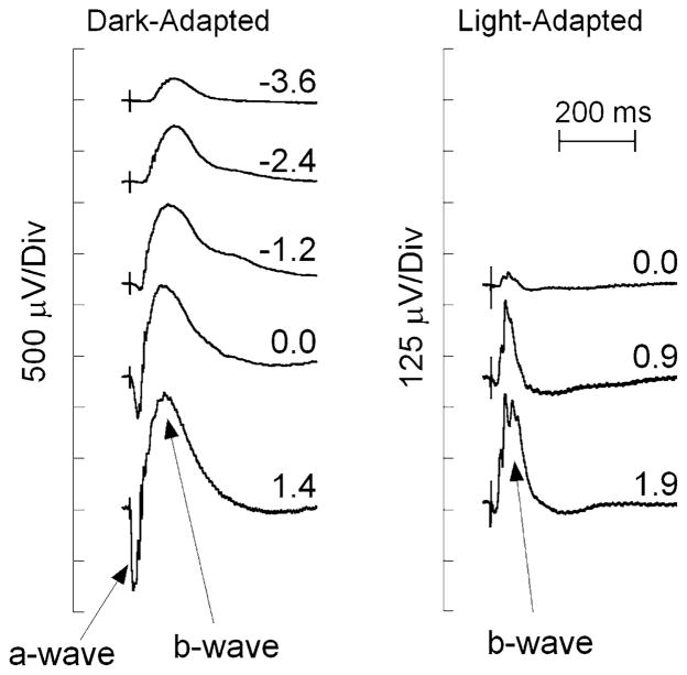
Fig. 2.
Representative ERGs recorded from the corneal surface of a WT mouse in response to strobe flash stimuli presented to the dark-adapted (left) or light-adapted eye (right). The b-wave is seen as a cornea positive potential, which increases in amplitude with increasing flash strength, indicated by the values next to each waveform, in log cd s/m2

