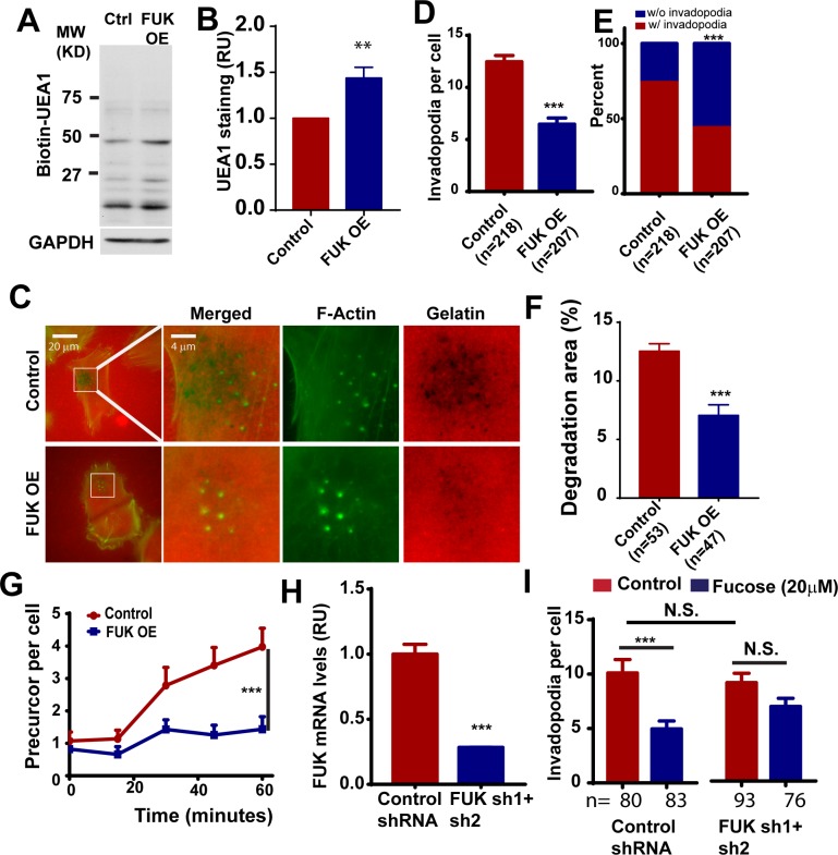
Fig 2. The ectopic expression of FUK in melanoma cells inhibits invadopodium formation and delays invadopodial initiation.
A, the effect of FUK overexpression on the fucosylation of WM793 cell surface proteoglycans. The plasma membrane proteins from control and FUK OE WM793 cells were separated using SDS-PAGE and detected by biotin-UEA1. B, the effect of FUK overexpression on cell surface fucosylation as detected by UEA1 staining and flow cytometry. C, representative images showing the effects of ectopically expressed FUK on invadopodium formation and TexasRed gelatin degradation in WM793. D and E, quantitation of the effect of ectopically expressed FUK on the invadopodium number per cell (D) and the proportion of invadopodia positive cells (E) in WM793. F, quantitation of the effects of FUK overexpression on gelatin degradation. G, the effects of ectopic FUK on the initiation of the assembly of invadopodia induced by 10% FBS. H, qPCR assay showing the effects of FUK sh1+sh2 on the mRNA transcript levels of FUK in WM793 cells. I, quantitation of the effects of FUK knockdown on fucose-mediated inhibition of invadopodium formation in WM793 cells. *** indicates p<0.001, as determined by two-tailed, two sample t-test (D, G, H, I) or two-tailed Fisher’s exact test (E). Numbers in parentheses indicates the number of cells used in quantitation. Representative results from at least three independent replicates were presented.

