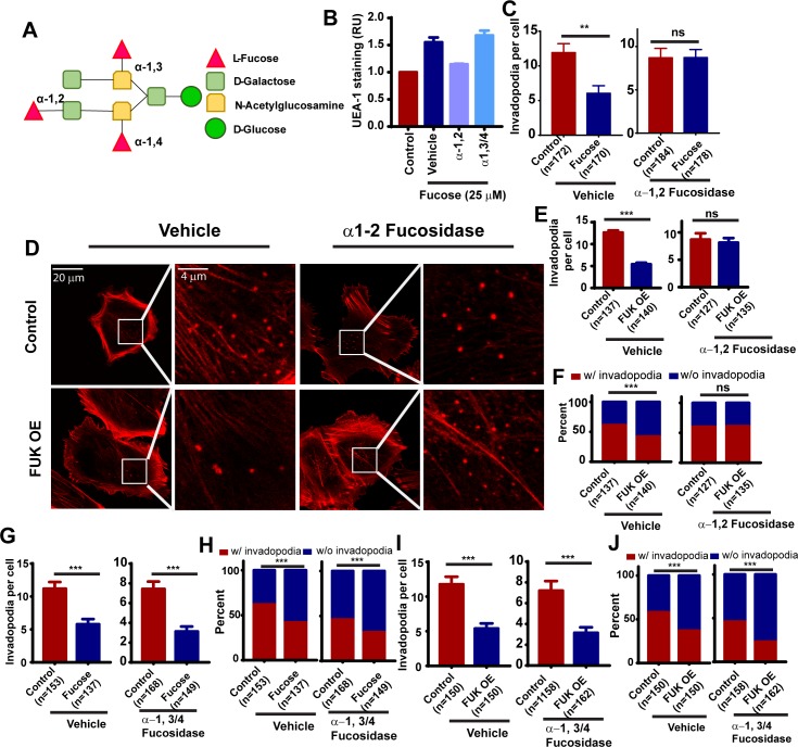
Fig 3. The fucose salvage pathway inhibits invadopodia through α1–2 fucosylation.
A, schematic illustration showing the modification of glycans by α-1,2, α-1,3 and α-1,4 branched fucosylations. B, the effects of L-fucose and fucosidase treatment on cell surface α1–2 fucosylation, as determined by UEA-1 staining and flow cytometry. C, quantification of the effects of α-1,2 fucosidase treatment on fucose-mediated inhibition of invadopodium formation in WM793 cells. D, representative invadopodium assay images showing the effect of α-1,2 fucosidase treatment on FUK-mediated inhibition of invadopodium formation in WM793 cells. E and F, quantitation of the effect of α-1,2 fucosidase treatment on the invadopodium number per cell (E) and the proportion of invadopodia positive cells (F) in control or FUK OE WM793. G and H, quantitation of the effect of α-1,3/4 fucosidase treatment on the average invadopodium number per cell (G) and proportion of invadopodia positive cells (H) in control or fucose-treated WM793. I and J, quantitation of the effect of α-1,3/4 fucosidase treatment on the average invadopodium number per cell (I) and proportion of invadopodia positive cells (J) in control or FUK-overexpressed WM793. ** and *** indicates p<0.01 and 0.001, respectively, as determined by two-tailed, two sample t-test (C, E) or two-tailed Fisher’s exact test (F, G). ns, not statistically significant. Numbers in parentheses indicates the number of cells used in quantitation. Representative results from at least three independent replicates were presented.

