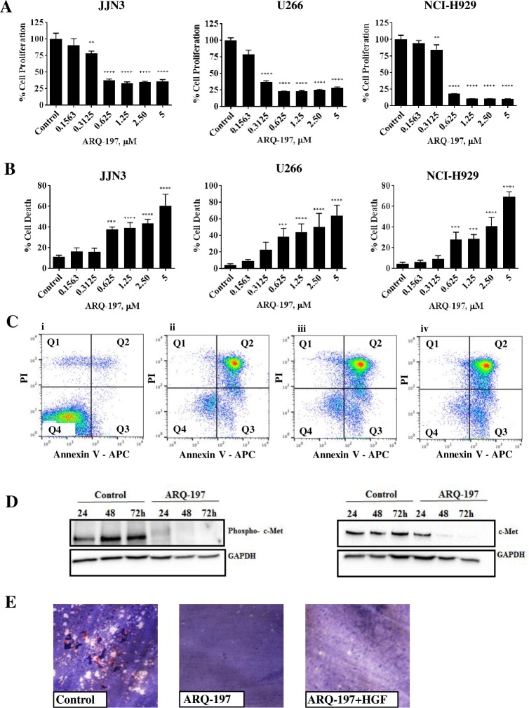
Fig 2. ARQ-197 inhibits cell proliferation, induces cell death by necrosis and reduces protein expression of phospho-c-Met and c-Met.
(A) JJN3, U266 and NCI-H929 cells were incubated with control media containing DMSO or ARQ-197 at 0.1563, 0.3125, 0.625, 1.25, 2.50 or 5 μM for 48 h. Cell proliferation was measured compared to DMSO control. (B) JJN3, U266 and NCI-H929 cells were incubated with control media containing DMSO or ARQ-197 at 0.1563, 0.3125, 0.625, 1.25, 2.50 or 5 μM for 48 h and counted using trypan blue exclusion and the percentage cell death calculated. All data displayed as mean ± SD and analysed using a normal one-way ANOVA, where significance is indicated by **P<0.01, ***P<0.001 or ****P<0.0001. (C) JJN3 cells were treated with control media containing DMSO (i) or ARQ-197 at 0.3125 (ii), 1.25 (iii) or 5 μM (iv) and then stained with annexin V/PI before flow cytometric analysis. In the flow cytrometry plots, Q1 relates to dead cells, Q2 necrotic, Q3 apoptotic and Q4 viable cells. (D) JJN3 cells were treated with control media containing DMSO or 1 μM ARQ-197 for 24 h, 48 h and 72 h and immunoblotted with an anti-phospho-c-Met antibody and an anti-c-Met antibody. (E) Representative images of osteoclasts treated with either control media containing DMSO, ARQ-197 1 μM or ARQ-197 1 μM with HGF 50 ng.

