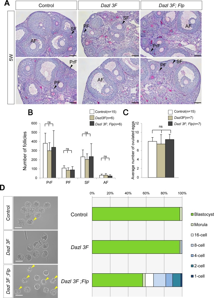
Fig 5. Defective pre-implantation development is a cause of the litter size reduction.
(A) PAS staining of control, Dazl 3F, and Dazl 3F;Flp ovaries at 5 weeks after birth. PrF, PF, SF, and AF are same as in Fig 2C. Scale bars, 100μm. (B) Follicle counting analysis for control (n = 15), Dazl 3F (n = 6), and Dazl 3F;Flp (n = 6) using ovarian sections at 5 weeks after birth. Error bars represent S.D. ns, no significant difference among control, Dazl 3F, and Dazl 3F;Flp. (C) The average number of ovulated eggs from control (n = 15), Dazl 3F (n = 7), and Dazl 3F;Flp (n = 7) females. Error bars, S.D. ns, no significant difference among control, Dazl 3F, and Dazl 3F;Flp (Tukey HSD test). (D) Analysis of pre-implantation development in BAC transgenic females. (Left) E3.5 embryos from control, Dazl 3F, and Dazl 3F;Flp females. Yellow arrowheads indicate abnormal embryos. Scale bars, 100μm. (Right) Proportion of embryos that developed to each stage up to blastocysts. Embryos collected from pregnant females at E3.5 were counted. Delayed embryos were cultured for an additional two days and ones that reached the blastocyst stage were added. Control (n = 75), Dazl 3F (n = 69) and Dazl 3F;Flp (n = 56).

