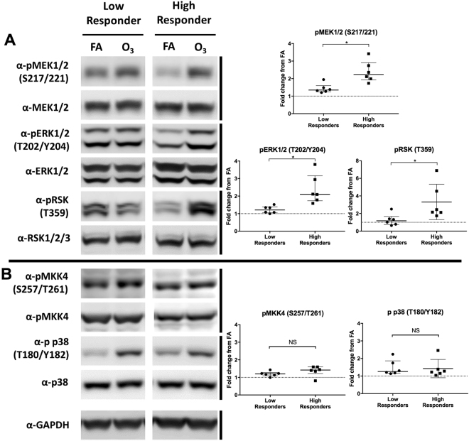
Figure 4.
The activation of ERK1/2, p38, and associated kinases in high and low-responding phBEC cultures. Cells were considered “high responders” if their IL-8 induction was above the group mean (4.2 fold change) and low responders were below this group mean. Protein was collected from twelve donors (n = 12), six high and six low responders. (A) Blots from two representative donors (one high, one low) show the phosphorylation of ERK1/2 and an upstream (MEK1/2) and a downstream (RSK) kinase, as well as p38 and its upstream kinase (MKK4). Both sets of blots were run on the same gel, immunoblotted, and imaged at the same time. (B) Densitometry analysis was used to calculate the fold change in activation (normalized to filtered air control) for each donor. Median activation and interquartile range are shown for each group. Differences between high and low responders were determined via Mann-Whitney test. *p < 0.05.

