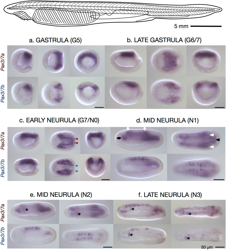
Figure 3.
Expression of Pax3/7a and Pax3/7b in a B. lanceolatum early developmental time course. Top: Illustrative line drawing of adult B. lanceolatum. Scale bar ≈5 mm. Below: Whole mount in situ hybridisation images of Pax3/7a-specific probe (top row of each block) and Pax3/7b-specific probe (bottom row of each block) in B. lanceolatum embryos. Views are presented, in left-to-right order: lateral, dorsal, and blastoporal (gastrula and early neurula only). Lateral and dorsal views are oriented with the anterior to the left. (a) Gastrula, G5, 10 hours post fertilisation (hpf). (b) Late gastrula, G6/7, 12 hpf. (c) Early neurula, G7/N0, 14 hpf. (d,e) Mid neurulae: N1, 16 hpf and N2, 21 hpf. (f) Late neurula, N3, 24 hpf. Domains of expression are marked throughout as follows: coloured arrowheads — differentially patterned neural plate border expression; black arrow — expression in the anterior mesodermal tissue; white arrows — in the anterior end and posterior of the neural tube; white arrowheads — in the postero-lateral somitic tissue; black arrowhead — in the postero-medial notochord tissue; asterisk — (placed immediately posteriorly to) the sinistral domain of expression found in both paralogues and in A. lucayanum. Scale bars = 50 micrometres.

