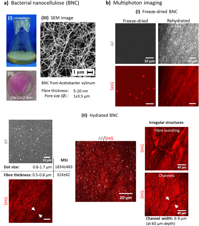
Figure 1.
BNC characterisation. (a) BNC biomaterial is formed as fleeces during static culture at the air-liquid interface (I). BNC fleece patch used for cell seeding (II). (III) SEM image (magnification M = 3,000x)22 providing insight into the nanocellulose network ultra-structure. Images I and III were provided by D. Kralisch. (b) Multiphoton pattern of dried BNC. The pattern of rolled BNC rehydrated with PBS dramatically changes within minutes. (c) Simultaneous AF/SHG images from within the BNC exhibit both dot- to elliptic-shaped AF signals and fibre-shaped SHG signals with different signal strengths (MSI, mean signal intensity). Representative overlay image shown in the middle. Irregular structures within the cellulose network were observed as well.

