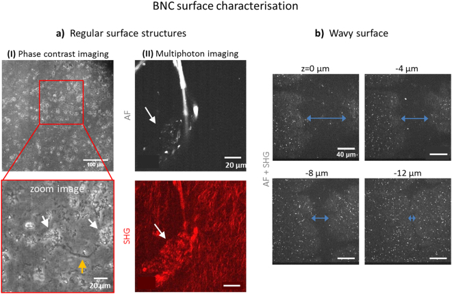
Figure 2.
Micro-structure of the BNC surface. In (a), regular surface structures are presented. (I) Phase contrast images (M = 10x) reveal structures reminding of cracks (orange arrow) and cavities (~10–20 µm in diameter, white arrows) all over the surface. In (II), a typical structural islet with an agglomerate multiphoton pattern is shown (white arrows in AF and SHG images). In (b), a trench is displayed in various depths which is large enough to act as cell seeding core (diameter of > 60 µm at surface, unseparated multiphoton image, M = 40x).

