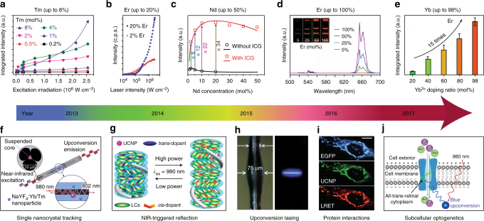
Fig. 2.
Selected milestones overcoming concentration quenching in homogeneously doped upconversion nanocrystals. a Integrated upconversion luminescence intensity as a function of excitation irradiance for a series of Tm3+-doped (0.2–8%) nanocrystals. Adapted from ref. 17. b Upconversion luminescence intensity of single 8 nm UCNPs with 20 and 2% Er3+, each with 20% Yb3+, plotted as a function of excitation intensity. Adapted from ref. 18. c Experimental results (black circle and red square) and theoretical modelling (black and red curves) of integrated upconversion luminescence intensities of a set of NaYF4:Nd3+ UCNPs with and without indocyanine green (ICG) sensitization. Adapted with permission from ref. 45 Copyright (2016) American Chemical Society. d Luminescence spectra of colloidal dispersion of NaYF4:x%Er@NaLuF4 nanocrystals (x = 5, 25, 50, 100); Inset: luminescence images of NaYF4:x%Er@NaLuF4 in cyclohexane excited with a 980 nm laser. Adapted with permission from ref. 25 Copyright (2017) American Chemical Society. e Integrated upconversion luminescence intensity of α-NaY0.98-xYbxF4:2%Er@CaF2 (x = 0.2, 0.4, 0.6, 0.8, 0.98). Adapted with permission from ref. 47 Copyright (2017) Royal Society of Chemistry. f Schematic of the experimental configuration for capturing upconversion luminescence of NaYF4:Yb3+,Tm3+ nanocrystals using a suspended-core microstructured optical-fibre dip sensor. Adapted from ref. 53. g Upon irradiation by a NIR laser at the high-power density, the reflection wavelength of the photonic superstructure red-shifted, whereas its reverse process occurs upon irradiation by the same laser but with the lower-power density. Adapted with permission from ref. 55 Copyright (2014) American Chemical Society. h Photographs of a microresonator with and without optical excitation. Adapted from ref. 56. i UCNPs functionalized with a nanobody recognizing enhanced green fluorescent protein (EGFP) could rapidly and specifically target to EGFP-tagged fusion proteins in the mitochondrial outer membrane, and this protein interaction process could be detected by lanthanide resonance energy transfer (LRET) in living cells. Scale bar: 10 μm. Reproduced with permission from Drees et al.57 copyright John Wiley and Sons. j Schematic of channelrhodopsin-2 activated in HeLa cells by strong blue upconversion luminescence from NaYbF4:Tm3+@NaYF4 core@shell structure. Adapted with permission from ref. 58 Copyright (2017) American Chemical Society

