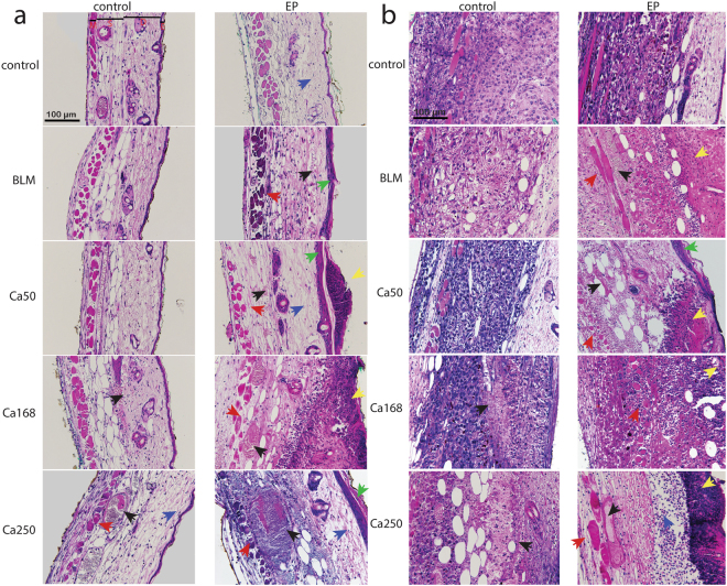
Figure 7.
Tissue damage in DWC after CaEP or ECT with bleomycin (BLM). Samples of two mice per treated group were subjected to histological determination of tissue damage. (a) Representative images of normal tissue sections that were exposed to the treatment and stained with haematoxylin and eosin. E epidermis, D dermis with hair follicles, S subcutaneous tissues with blood vessels, M striated muscle layer. (b) Representative images of tumour sections exposed to the treatment. Blue arrow – oedema; black arrow – blood vessels and extravasation; green arrow – damaged epidermis; red arrow – muscle necrosis; yellow arrow – tissue necrosis.

