
FIGURE 1.
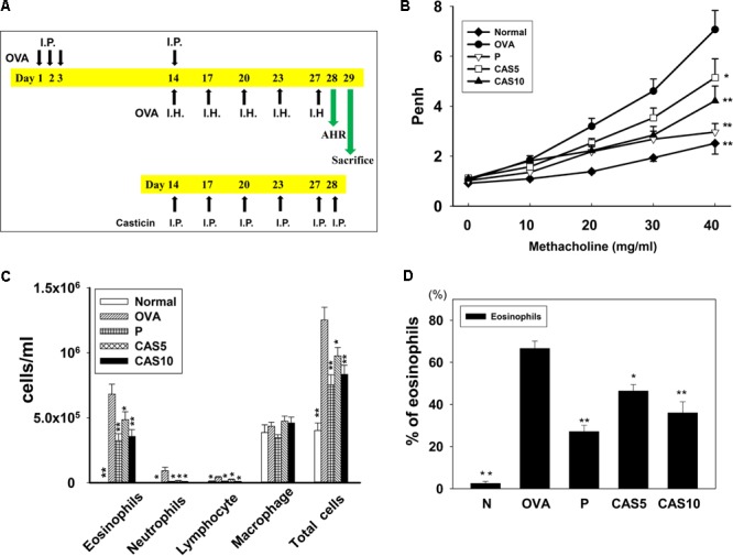
Effect of casticin on AHR and cell counts in BALF from asthmatic mice. (A) On days 1–3 and 14, mice sensitized with OVA by intraperitoneal injection (I.P.) and challenged with 2% OVA inhalation (I.H.) on days 14, 17, 20, 23, and 27. One hour before the OVA challenge or methacholine inhalation, mice were treated with I.P. casticin or prednisolone. (B) AHR (Penh values) was measured via inhalation of increasing methacholine doses (0–40 mg/ml). (C) Inflammatory cells and total cells were measured in BALF and (D) presented the percentage of eosinophils in BALF. The data are presented as means ± SEM of three independent experiments (n = 12). ∗p < 0.05, ∗∗p < 0.01 compared to the OVA control group. Mice were divided into normal (N), OVA-sensitized mice (OVA), prednisolone control (P), and casticin treatment (CAS5 and CAS10) groups.