
FIGURE 2.
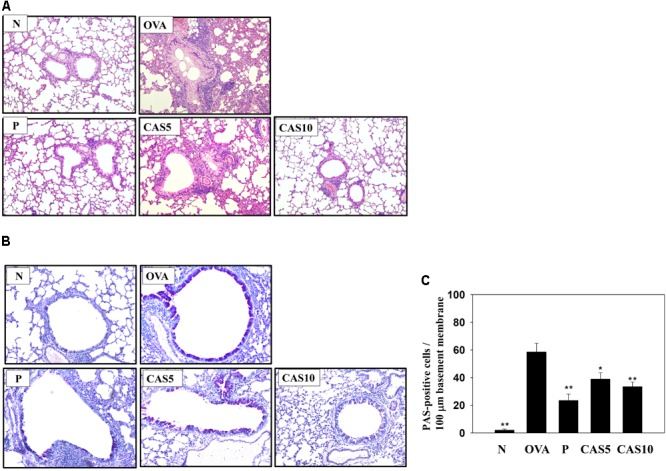
Effects of casticin (CAS) on asthmatic lung tissue. Histological sections of lung tissues from normal (N) and OVA-stimulated (OVA) mice with or without prednisolone (P) and CAS treatment. (A) CAS reduced eosinophil infiltration (H&E stain; 200× magnification). (B) Periodic acid-Schiff (PAS)-stained lung sections show goblet cell hyperplasia (200× magnification). (C) Results are expressed as the number of PAS-positive cells per 100 μm of basement membrane. The data are presented as means ± SEM of three independent experiments (n = 6). ∗p < 0.05, ∗∗p < 0.01 compared to the OVA control group.