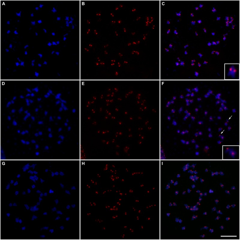
FIGURE 5.

Fluorescent in situ hybridization of centromeric probes on meiotic cells at diakinesis: (A) Chromosomes of “Caiana Fita” (Saccharum officinarum) stained with DAPI (blue); (B) Centromeric sites hybridized with the CENT probe detected with anti-DIG-rhodamine (red); (C) Superposition of the images (A/B) showing 40 bivalents; the inset shows a typical bivalent; (D,G) Chromosomes of “IACSP93-3046 stained with DAPI (blue); (E,H) Centromeric sites hybridized with the CENT probe detected with anti-DIG-rhodamine (red); (F) Superposition of the images (D/E) showing 56 bivalents plus two univalents; arrows point to the univalents and the inset shows a univalent and a bivalent; (I) Superposition of the images (G/H) showing 56 bivalents. Bar, 10 μm.
