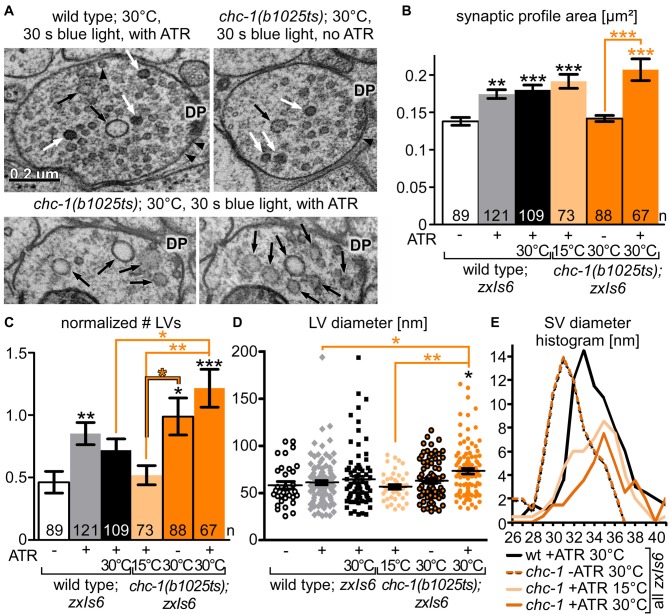
Figure 8.
Ultrastructural analysis of chc-1 mutants demonstrates a role in synaptic endosome breakdown during SV recycling. (A) Example electron micrographs of wild-type and chc-1(b1025ts) mutant synapses, photostimulated for 30 s under non-permissive temperature (30°C), in animals raised without or with ATR. Structures are indicated as in Figure 6A. (B) Synaptic dimension (area) for the indicated genotype and conditions, for the indicated number of profiles, analyzed as in Figure 6B. Two to three animals and 11–19 synapses, with 67–121 profiles were analyzed for each condition (see Supplementary Table S1). (C) LV numbers observed per profile, normalized to a typical synaptic profile area, and averaged across the indicated number of profiles. (D) Individual LV diameters, shown as scatter plot with means and SEM. Statistical analysis: one-way ANOVA with Tukey correction in (B–D); ***p < 0.001; **p < 0.01; *p < 0.05. (E) SV diameter distribution, as a histogram, filtered with a sliding average across the neighboring two values.

