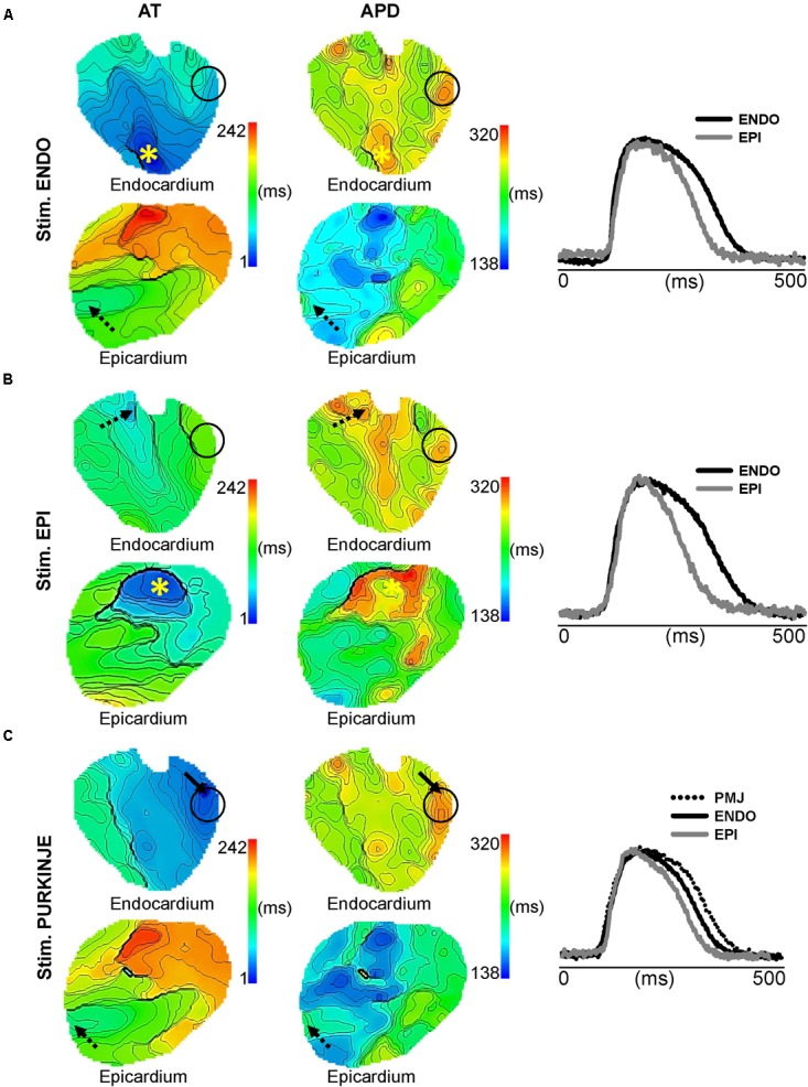
FIGURE 4.

Impact of ischemia on transmural APD heterogeneity. Endocardial and epicardial AT and APD maps and corresponding AP traces as shown in Figure 2 for ENDO (A), EPI (B), and PF (C) pacing. Stimulation sites are indicated by (∗), arrows show origins of early activation for PF stimulation in C, dashed arrows indicate origin of breakthrough, and black circles show PMJs location on AT and APD maps for all pacing sites. Isochrones are 5 ms spacing for endocardial maps and 10 ms for epicardial maps.
