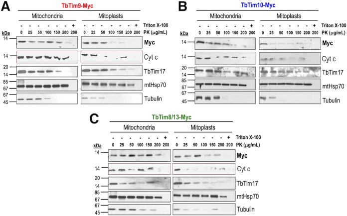
FIG 3 .
Submitochondrial localization of the small TbTim proteins. Proteinase K protection assays of mitochondria (left panels) and mitoplasts (right panels) isolated from T. brucei cells expressing (A) TbTim9-Myc, (B) TbTim10-Myc, or (C) TbTim8/13-Myc were performed. Mitochondria and mitoplasts were treated with various concentrations (0 to 200 µg/ml) of PK as described in Materials and Methods. Proteins were analyzed by SDS-PAGE and immunoblot analysis using anti-myc, anti-Cyt c, anti-TbTim17, anti-mtHsp70, and anti-tubulin antibodies.

