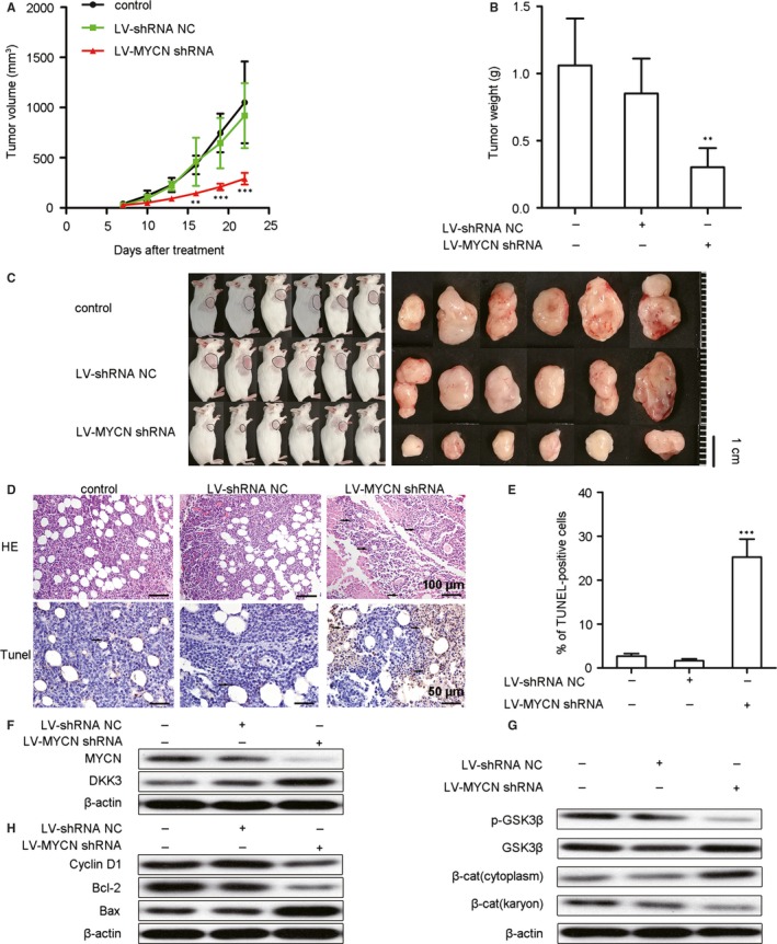
Figure 5.

MYCN shRNA suppresses tumour growth via the restoration of DKK3‐mediated inhibition of the Wnt/β‐catenin pathway in a murine Nalm6 xenograft model. Mice burdened with growing Nalm6 tumours were infected with Lv‐shRNA NC or Lv‐MYCN shRNA or left untreated (control), and the tumour sizes (A) were measured for 22 days (n = 6). The tumour weights (B) were also measured after 22 days (n = 6). Images of tumour‐bearing mice (C) show that the tumours were smaller in MYCN shRNA‐treated animals. (D) Haematoxylin and eosin (H&E) (upper) and TUNEL (lower) staining of the xenograft tumour tissues from 3 mice of each group. In the H&E‐stained sections, the original magnification was 200 × . In the TUNEL‐stained sections, positive cells are indicated by brown staining, with an original magnification of 400 × . Representative fields are shown. Arrows indicate tumour cells. (E) The TUNEL‐positive cells were measured from 3 randomly chosen fields (3 × 104 μm2/field; n = 9). (F) Representative Western blots show the protein expression of MYCN and DKK3. (G) Representative Western blots show the expression of p‐GSK3β, GSK3β and β‐catenin (cytoplasmic and nuclear). (H) Representative Western blots show the expression of Bax, Bcl‐2 and cyclin D1. Expression of proteins was measured by Western blotting, and β‐actin was used as a loading control; we used 3 mice from each group. All data are presented as the means ± SD. **P < .01 vs. Lv‐shRNA NC; ***P < .001 vs. Lv‐shRNA NC. NC: negative control, β‐cat: β‐catenin
