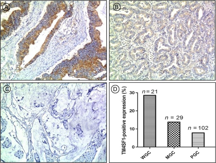
Figure 2.

Higher levels and strong expression rate of TM4SF1 were associated with GC tissues of higher‐grade differentiation. TM4SF1 expression in GC tissues was determined by immunohistochemtry. Expression rates of TM4SF1 (D) in (A) well, (B) moderately, and (C) poorly differentiated GC tissues.
