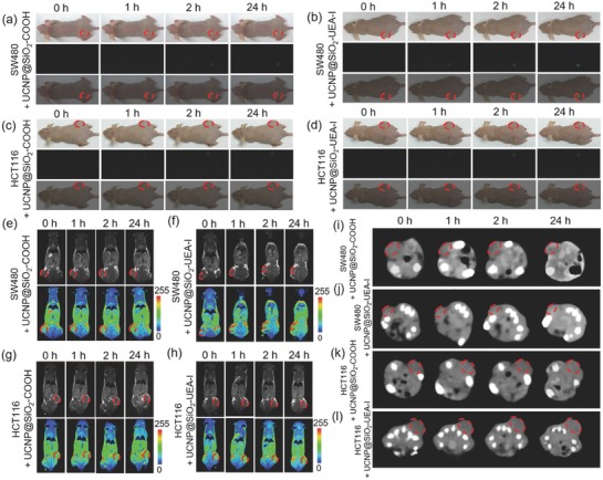
Figure 4.

In vivo a–d) UCL (from up to bottom: bright field mode, UCL mode (green channel), and merging mode), e–h) MR, and i–l) coronal CT images of SW480 tumor‐ and HCT116 tumor‐bearing nude mice after intravenous injection of UCNP@SiO2—COOH and UCNP@SiO2–UEA‐I at different timed intervals, respectively (the 0 h means preinjection).
