
Figure 1.
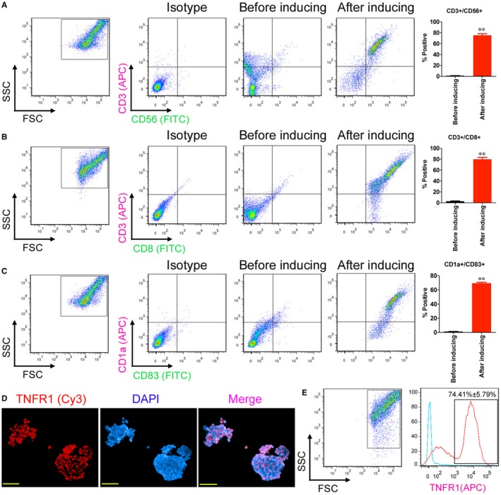
Induction of OCPMB‐derived mononuclear cells in vitro to differentiate into DC and CIK cells. A, FACS detection results showed that stimulated mononuclear cells significantly expressed high levels of the CIK markers CD3 and CD56. **P < .01 vs before induction; n = 6; B, FACS detection results showed that stimulated mononuclear cells significantly expressed high levels of the CIK makers CD3 and CD8. **P < .01 vs before induction; n = 6; C, The FACS detection results showed that the stimulated mononuclear cells significantly expressed high levels of the DC markers CD1a and CD83. **P < .01 vs before induction; n = 6; D, Immunofluorescence staining results showed that the enriched CD44+/CD133+ OCSCs expressed high levels of TNFR1 protein. Scale bar = 30 μm; E, The FACS detection results showed that the positive TNFR1 protein detection rate among the enriched CD44+/CD133+ OCSCs reached approximately 74.41%