
Figure 3.
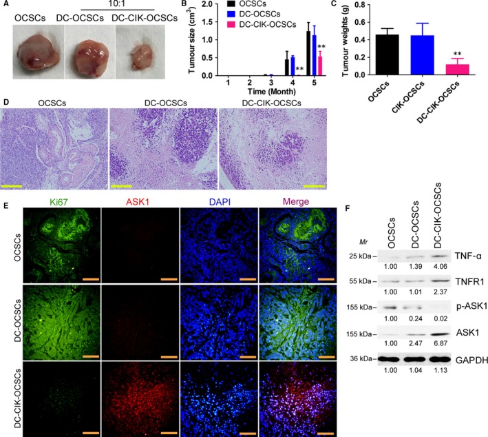
OCPMB‐derived DC‐CIK cells significantly inhibited the growth of CD44+/CD133+ OCSCs in nude mice. A, DC or DC‐CIK cells were mixed with OCSCs at a ratio of 10:1 and cultured in vitro for 24 h. The cells were collected and subcutaneously injected into nude mice. After 5 mo, tumour tissues derived from each group of cells were isolated; B, The volume of tumour tissues in the DC‐CIK‐OCSC group was significantly smaller than those in the simple OCSC group and the DC‐OCSC group. **P < .01 vs OCSCs; n = 6; C, The weight of tumour tissues in the DC‐CIK‐OCSC group was significantly lower than those in the simple OCSC group and the DC‐OCSC group. **P < .01 vs OCSCs; n = 6; D, The HE staining results showed that tumours in all groups met the characteristics of mixed‐type epithelial ovarian cancer. The tumour tissues in the DC‐CIK‐OCSC group had more necrotic cells, significantly reduced numbers of tumour cells and significantly reduced malignancy. Scale bar = 30 μm; E, The immunofluorescence staining results showed that the number of Ki67‐positive cells in tumour tissues from the DC‐CIK‐OCSC group was significantly lower than those in the other 2 groups, whereas the number of ASK1‐positive cells was significantly higher than those in the other 2 groups. Scale bar = 30 μm; F, The Western blot results suggested that the expression levels of TNF‐α, TNFR1 and ASK1 in tumour tissues in the DC‐CIK‐OCSC group were significantly higher than those in other groups, whereas the expression level of p‐ASK1 protein was significantly lower than those in other groups