
Figure 3.
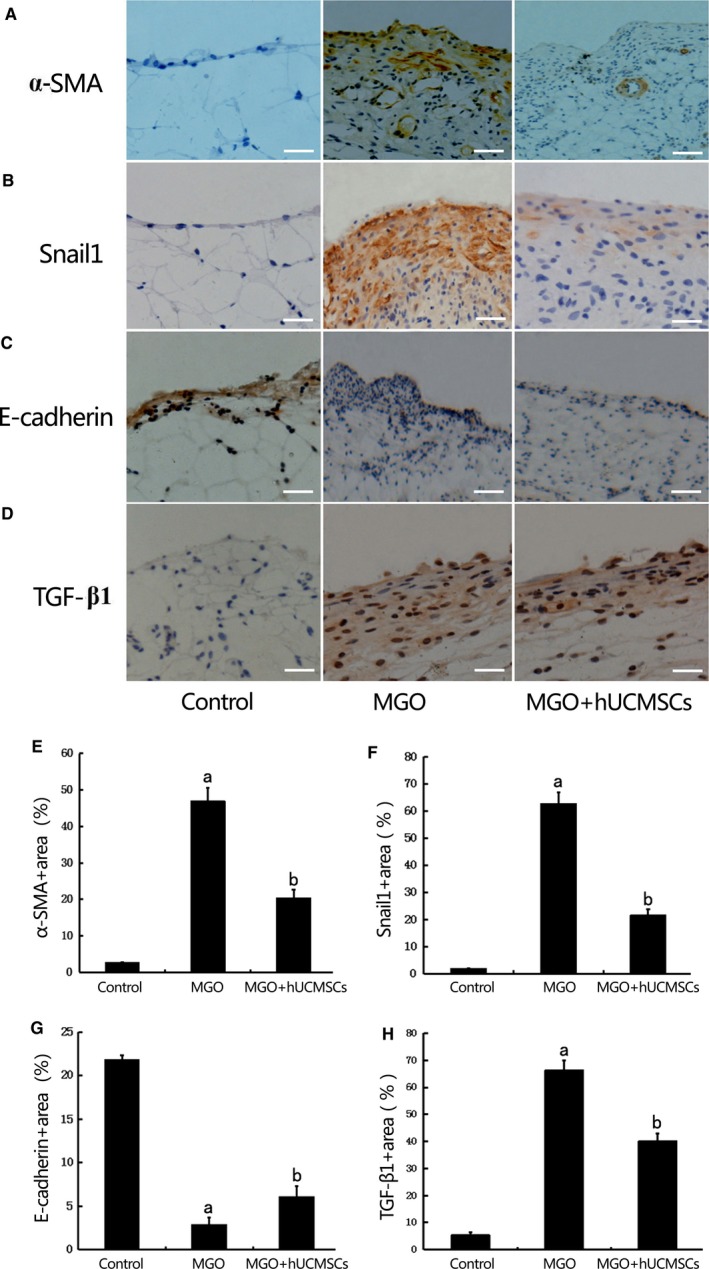
Effects of hUCMSCs on epithelial to mesenchymal transition (EMT) in the MGO‐induced peritoneal tissues in rats. The expression of α‐SMA (A), Snail1 (B), E‐cadherin (C) and TGF‐β1 (D) were detected by immunohistochemistry in peritoneal tissues of Group Control, Group MGO and Group MGO + hUCMSCs (×400 magnification, bar = 100 μm).(E‐H)Quantitative analysis revealed that α‐SMA, Snail1 and TGF‐β1 protein expression were significantly higher, and E‐cadherin was significantly lower in rats treated with MGO than that in control rats. hUCMSCs significantly down‐regulated α‐SMA, Snail1 and TGF‐β1 and up‐regulated E‐cadherin expression in MGO‐induced peritoneal fibrosis (PF) rats(n = 10‐12). Data were analysed by Student's t test and displayed as mean±SEM of 10‐12 rats per group. aP<0.05 vs Group Control, bP<0.05 vs Group MGO