
Figure 2.
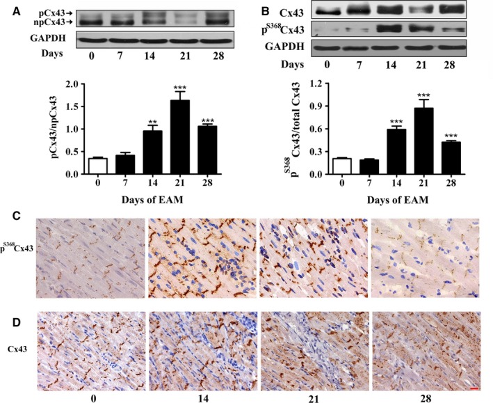
Experimental autoimmune myocarditis (EAM) induces up‐regulation of pS 368Cx43. A, The ratio between phosphorylated and non‐phosphorylated form of Cx43 in the cardiac ventricles on day 0, 14, 21 and 28 of EAM. Upper panel: Western blot; Lower panel: Statistical summary. B, Relative pS 368Cx43 level in the cardiac ventricles on day 0, 14, 21 and 28 of EAM (n = 6‐8). Upper panel: Western blot; Lower panel: Statistical summary. *P < .05 vs day 0; **P < .01 vs day 0; ***P < .001 vs day 0. C and D, Sections of stained pS 368Cx43 (C, brown) and Cx43 (D, brown) in the longitudinal sections of the inflammatory area of cardiac ventricles on day 0, 14, 21 and 28 of EAM. Bar = 20 μm