Figure 4.

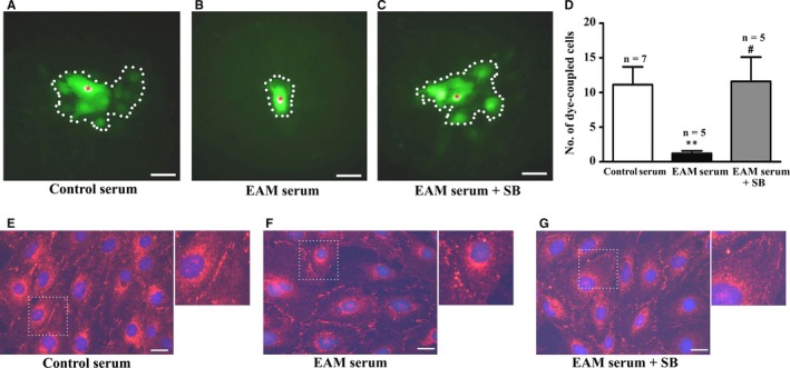
Experimental autoimmune myocarditis (EAM) serum suppresses cell‐to‐cell communication via p38 signalling. A‐C, Image of dye‐coupling between the cells treated with 0.25% serum from the control rats (A) and EAM (B) rats as well as 0.25% EAM serum in the presence of 10 μmol/L SB203580 (C). Dotted line: dye‐coupled cells. Red asterisk: dye‐injected cell. D, Statistical summary for dye coupled cells; *P < .01 vs control; # P < .05 vs EAM serum. E‐G, Immunofluorescent image of Cx43 in the cells treated with 0.25% serum from the control (E) and EAM (F) rats as well as 0.25% EAM serum in the presence of 10 μmol/L SB203580 (G). For greater clarity, areas in dotted squares are enlarged twofold in the upper right of each figure. Bar = 50 μm
