
Figure 5.
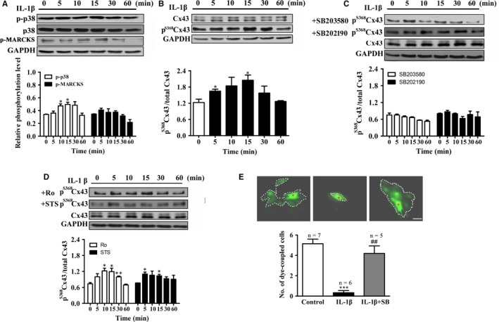
IL‐1β up‐regulates pS 368Cx43 level and suppresses cell‐to‐cell communication via p38 MAPK. A, Time course of 10 ng/mL IL‐1β on p‐p38 (n = 3). Upper panel: Western blot; Lower panel: Statistical summary. B‐D, Time course of 10 ng/mL IL‐1β on relative pS 368Cx43 (B), in the presence of p38 inhibitors (10 μmol/L SB203580 or SB202190) (n = 3) (C) or PKC inhibitors (2 μmol/L Ro‐32‐0432 or 100 nmol/L STS) (D). Upper panel: Western blot; Lower panel: Statistical summary. E, Effect of IL‐1β on the number of dye coupled cells in the absence or presence of 10 μmol/L SB203580. Dotted line: dye‐coupled cells. Red asterisk: dye‐injected cell. *P < .05 vs control; ***P < .001 vs control; ## P < .01 vs IL‐1β. Bar = 50 μm