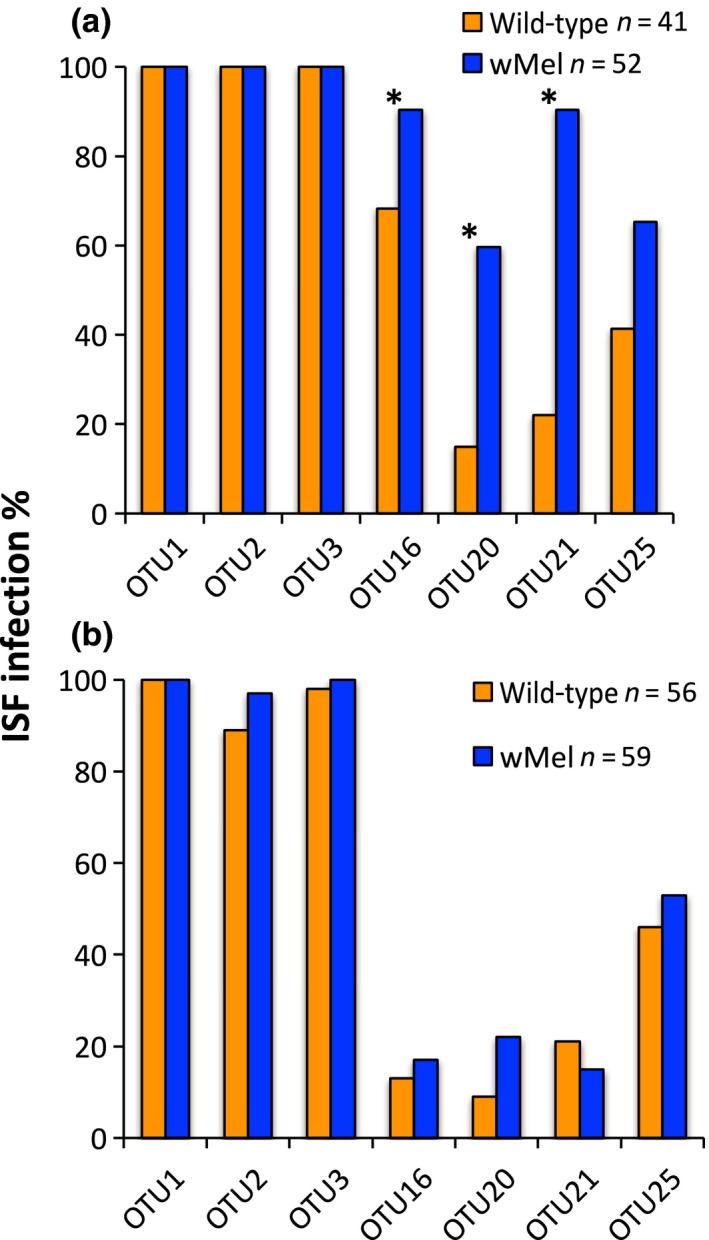
Figure 3.

ISF infection rates in mosquitoes for a subset of OTUs as determined by RT‐qPCR. (a) In the field, three of the OTUs were more common in wMel‐infected mosquitoes than WT (*p < .0125). (b) In the laboratory, there were no differences in ISF infection rates between wMel and wild‐type mosquitoes in the laboratory (p = .27)
