
Figure 1.
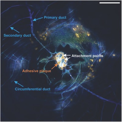
Confocal microscopy image of a barnacle after exposure to artificial seawater (ASW) containing a neutral lipid probe (Bodipy 493/503, green) and a primary amide probe (Alexa Fluor 647 NHS, orange). The image shows the cyprid attachment site (white arrows) and adhesive plaque (orange arrow), as well as part of the cementing apparatus that consists of primary ducts, secondary ducts, and circumferential ducts around the barnacle base. The cuticle and ducts exhibit strong autofluorescence on the blue channel (ex 405 nm). Scale bar represents 100 µm.