
Figure 2.
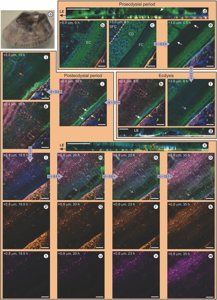
a) Photograph of an acorn barnacle. b–x) Confocal microscopy images of a barnacle in ASW containing a nucleic acid probe (DAPI, blue), a neutral lipid probe (Bodipy 493/503, green), a reactive carbonyl species probe (Alexa Fluor 555 hydrazide, magenta), and a primary amide probe (Alexa Fluor 647 NHS, orange). Distances in the top left corners correspond to height above the interface. All scale bars represent 20 µm. (b) Cuticle‐secreting epidermal cells (EC) anchored to the side shell at the leading edge (LE) of the barnacle by columnar cells. (c) Early stage of circumferential duct (CD) and folded cuticle (FC) development. Dashed line denotes the front of the base plate, which is surrounded by shell‐forming cells (SFC). (d) Orthogonal view of a 10 µm Z‐stack showing the forming FC (white arrow) above the old cuticle (gray arrow). (e) Lipid granules (white arrows) coalesce along the FC. (f) Older cuticle degrades and sheds during ecdysis (gray arrow), but remains intact and attached to the newly sclerotized cuticle (blue arrow). (g) Orthogonal view of a 20 µm Z‐stack showing the new cuticle (blue arrow) and molted cuticle (gray arrow). (h,i) Lipid secretions (white arrows) accumulate at the leading edge and are pulled outside as the barnacle expands. A nonlabeled zone is evident between the biofilm and the leading edge (yellow arrows). j,k) Change in fluorescence from green to orange indicates protein is accumulating within lipid granules; granules smear as barnacle grows. l–o) Molted cuticle tears (gray arrows) as the barnacle expands; fibrillar proteinaceous cement accumulates between cuticular layers. Secreted cement fibrils (p–s) gradually oxidize, presenting more reactive carbonyl groups over time (t–w). (x) Orthogonal view of an 8 µm Z‐stack shows epidermal cells secreting cement fibrils through unfolded sections of the new cuticle.