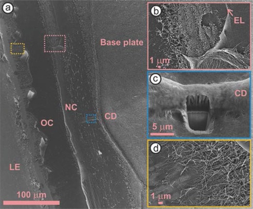
Figure 3.

a) Scanning electron microscopy image of a barnacle attached to a substrate, after its side shell was removed. b) A band of cement nanofibrils are present along the ecdysial line (EL), between the new (NC) and old cuticle (OC) layers. c) Focused ion beam milling exposes the interior of a circumferential duct (CD). d) Cement fibrils intermix with other barnacle deposits at the leading edge (LE) under the OC.
