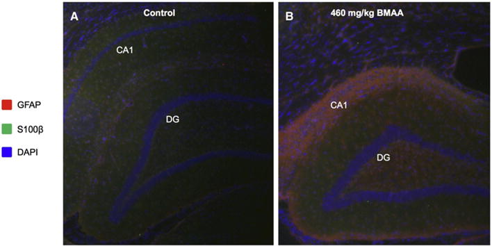
Fig. 4.

Immunohistochemistry reveals glial activation in the adult hippocampus upon neonatal BMAA exposure. Double antigen staining was performed against GFAP and S100β on hippocampal sections of control (A) and BMAA exposed animals (B). The data show massive gliosis and reactive astrocyte species localizing to the histopathologically altered region in the hippocampus of BMAA treated rats. This was indicated by increased GFAP immune-reactivity (red) in the CA1 and DG of BMAA exposed animals (B) compared to controls (A). GFAP positive cells were also found to stain positively for S100β serving as additional marker for astrocyte activation (green, B). Magnification: lens = ×10.
