Figure 2.
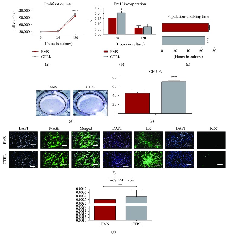
Growth kinetics of ASCs cultured in control conditions. Cell number estimated using a resazurin-based assay (a) and incorporation of BrdU (b). Population-doubling time was calculated using the number of cells in each of the experimental time points (c). The ability of cells to form colonies originating from one cell was evaluated by CFU-F assay. Representative photographs showing colonies stained with pararosaniline (d) and quantitative data obtained by the application of a CFU-F algorithm (e). The morphology of cells was investigated using fluorescence staining for f-actin and endoplasmic reticulum (ER) (f). Moreover, proliferation of cells was estimated by immunofluorescence staining for the Ki67 antigen as presented on representative images. Data was quantified and the Ki67/DAPI ratio was calculated (g). Results are expressed as mean ± SD. Scale bar 100 μm. ∗p < 0.05, ∗∗p < 0.01, and ∗∗∗p < 0.001.

