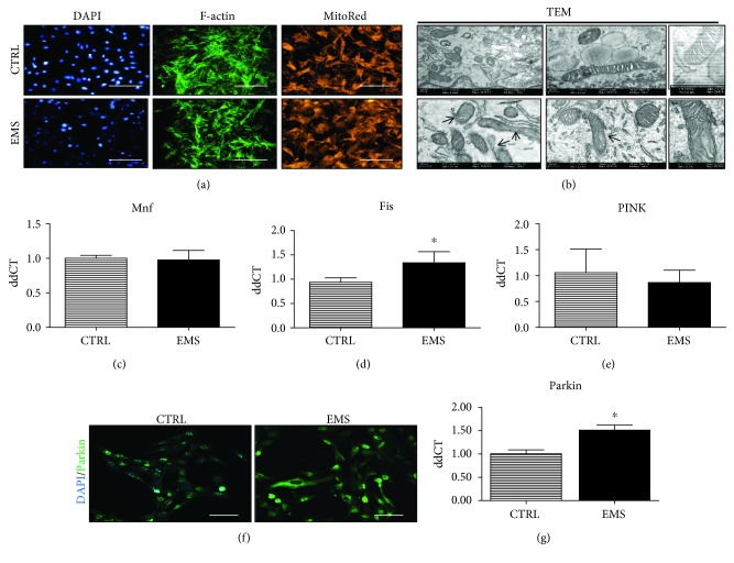
Figure 7.
Mitochondrial dynamics and clearance in ASCCTRL and ASCEMS cultured under adipogenic conditions. Using fluorescence staining, cells' cytoskeleton (f-actin) and mitochondrial net were visualized (a). Moreover, a TEM microscope allowed for a deeper assessment of mitochondrial morphology (b). Mitochondria from ASCCTRL presented typical, elongated morphology, while ASCEMS cells were characterized by mitochondrial aberrations including membrane and cristae raptures. Moreover, mitochondrial dynamics was assessed by RT-PCR as the expression of Mnf (c) and Fis (d) genes was investigated. However, no differences were observed between groups. Similarly, no differences in PINK expression were noted (e). To evaluate the amount of Parkin protein, immunofluorescence staining and RT-PCR were performed. Representative photographs obtained from a confocal microscope indicated increased Parkin accumulation in ASCEMS (f). Those data were also confirmed by the analysis of Parkin mRNA levels, as it was significantly upregulated in ASCEMS (g). Results are expressed as mean ± SD. Scale bar 250 μm. ∗p < 0.05.

