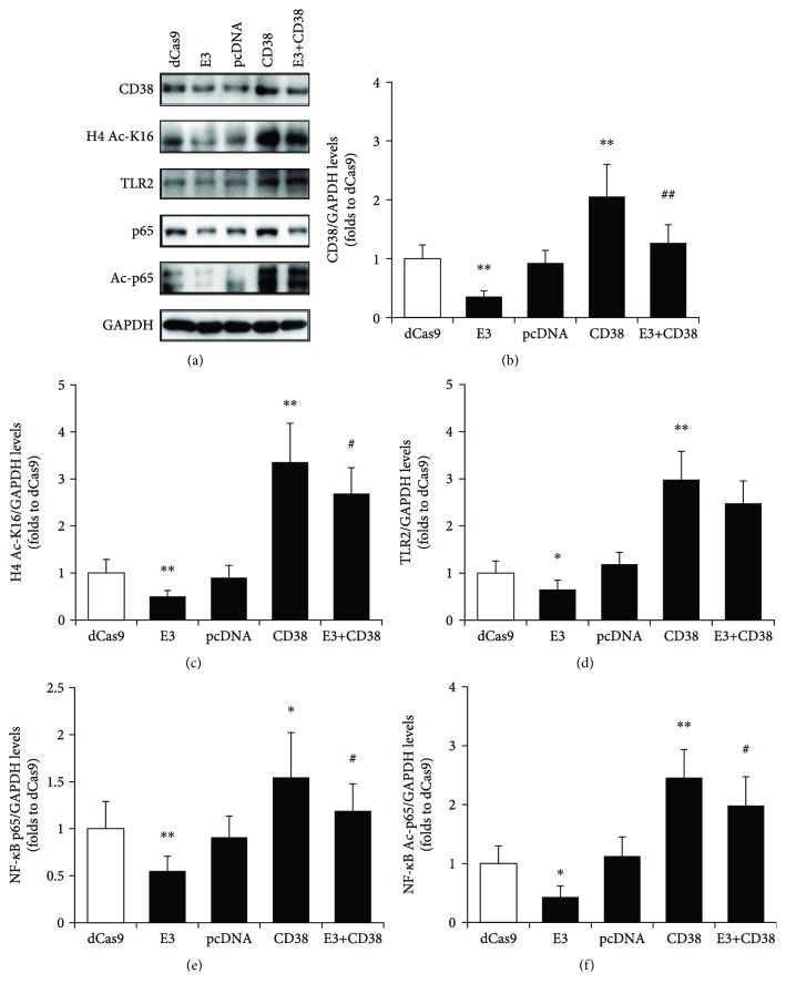
Figure 5.
Re-expression of CD38 in RAW264.7 cells reverses Sirt1/NF-κB/TLR2 signaling. The different clones of RAW264.7 cells, including the negative control dCas9 and pcDNA, the CD38 knockdown cell line E3, the overexpression clone of CD38, and the line co-transfected with E3 and CD38 (E3+CD38), were selected for the experiment. The expressions of CD38, acetylated histone 4 lysine 16 residue (H4 Ac-K16), TLR2, NF-κB p65, and acetyl-p65 (Ac-p65) proteins were detected by Western blot (a) and quantitatively determined for CD38 (b), H4 Ac-K16 (c), TLR2 (d), NF-κB p65 (e), and NF-κB Ac-p65 (f). The levels of H4 Ac-K16 represent the activity of Sirt1. GAPDH was used as an internal control. The data represent the mean ± S.D. from three independent experiments. ∗ p < 0.05 and ∗∗ p < 0.01 compared with the dCas9 group; # p < 0.05 and ## p < 0.01 compared with the CD38 group.

