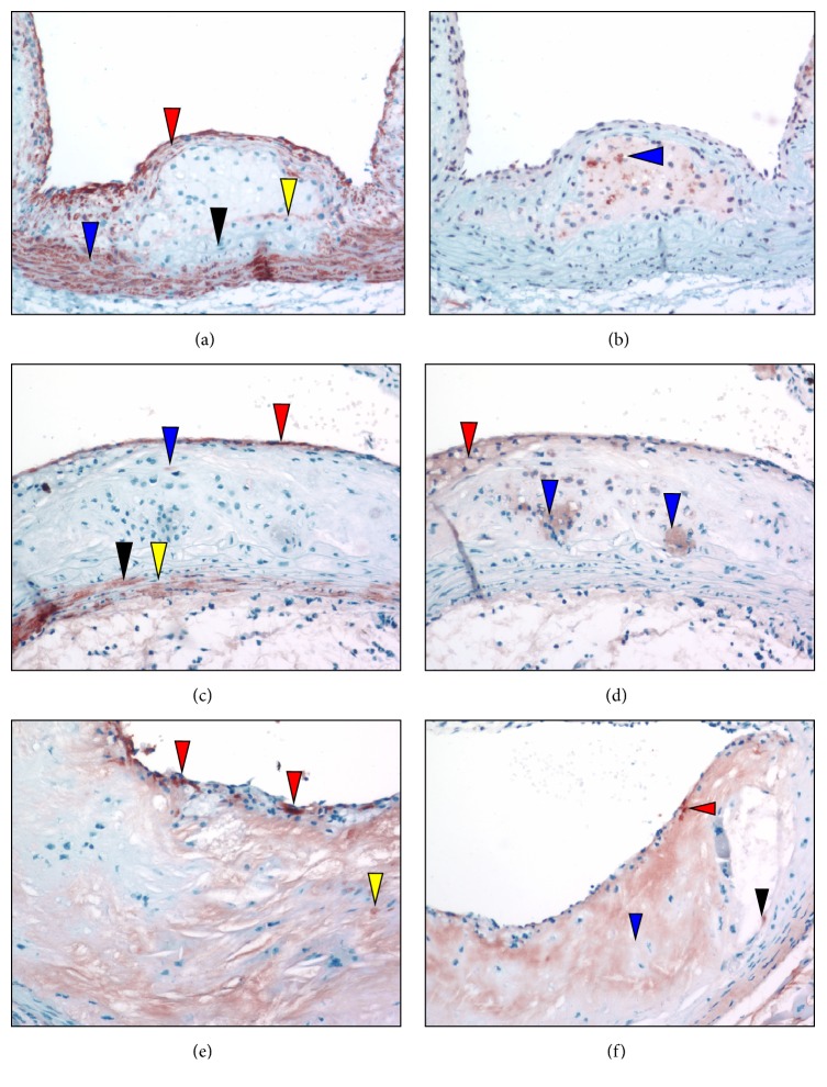
Figure 10.
Anti-SMA and anti-SMemb immunostaining positive foci of aortic sinuses from typical L−/−/A−/− mouse at various ages. (a) 24 weeks of age. Anti-SMA immunostaining reveals a cellular multilayered SMC α-actin positive region associated with the fibrous cap (red arrowhead), as well as the normally positive medial compartment (blue arrowhead). However, at the base of the lipid core, within the media, an area devoid of positive cells is evident (black arrowhead) with a faintly positive region immediately underlying the core (yellow arrowhead). (b) 24 weeks of age. Several SMemb-positive foci are identified in the core (blue arrowhead), in an area devoid of SMA positive cells. (c) 36 weeks of age. Anti-SMA immunostains indicate numerous single layered positive cells (red stain) at the endothelium. They consist of weak to highly positive SMA foci among other negative cells. Only one weakly positive cell is located under the subcapsular region (blue arrowhead). The medial compartments of the arterial wall are populated with both SMA positive (black arrowhead) and negative (yellow arrowhead) cells. The inner medial compartment (below the lipid core) appears distended and is mainly negative for SMA. (d) 36 weeks of age. Several diffuse SMemb-positive foci are identified within the broad end of the foam cell-containing cap (red arrowhead). Additional foam cell clusters within the core are also positive (blue arrowheads). These areas within the core were negative for SMA. (e) 48 weeks of age. Anti-SMA immunostains disclose SMC-actin positive (red) cells in the subluminal region, which are intensely positive (red arrowheads) and diffusely scattered among SMA negative border cells. A few positive cells are identified within the core (yellow arrowhead). (f) 60 weeks of age. Anti-SMA immunostains show SMA positive cells that are faintly visible in the subendothelium (red arrowhead) with a mostly acellular necrotic core containing few positive cells (blue arrowhead). A positive cell is identified at the base of the fragmented lesion within the core (black arrowhead). Original magnification, 100X.

