Figure 9.
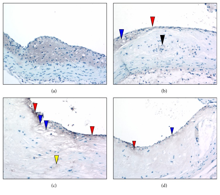
Antimacrophage immunostaining of aortic sinuses from typical L−/−/A−/− mouse at various ages. (a) 24 weeks of age. Antimacrophage stains demonstrate that the foam cell core (gray area) consists predominantly of macrophages. (b) 36 weeks of age. Antimacrophage immunostains show that the cap now consists of scattered macrophages (red arrowhead). At the broadest area of the cap, the macrophages appear as foam cells adjacent to the core (blue arrowhead). There are a few lightly stained (gray) positive stains within the core (black arrowhead). (c) 48 weeks of age. Antimacrophage immunostaining shows macrophages (gray stains) within the thinned cap in the subendothelium (red arrowheads). Some are associated with breakpoints of the endothelium underlying some faintly positive foam cells (blue arrowheads). The central core shows focal but scattered areas of faintly stained cells (yellow arrowhead). (d) 60 weeks of age. Antimacrophage immunostaining reveals small clusters of macrophages within the endothelial (blue arrowhead) and subendothelial areas (red arrowhead). Original magnification, 100X.

