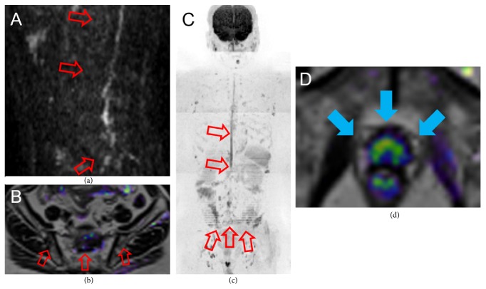
Figure 3.
WB-MRI at the time of CRPC diagnosis. Sagittal DWI of the spine (a), coronal DWI + T2 fusion (b), and whole-body DWI (c) showed few high signals. Coronal DWI + T2 fusion of the pelvis (d) revealed diffuse high signal in the prostate. WB-MRI, whole-body magnetic resonance imaging; CRPC, castration-resistant prostate cancer; DWI, diffusion-weighted imaging.

