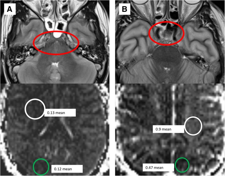
Fig. 4.

Correlation of hypointensity on b50 ADC maps in the BD population in comparison with flow voids. T2w (upper row) and b50 ADC (lower row) MRI images in two subjects with BD, with a subject A demonstrating loss of flow voids, and b subject B demonstrating maintained flow voids, at the skull base (red circles). Subject A demonstrated lower white matter (white circle) b50 ADC values than the white matter (white circle) in subject B (0.13 vs 0.92 × 10⁻³ mm2/s). The gray matter (green circles) was markedly low in both subjects, although lower in the subject without flow voids (0.12 vs. 0.47 × 10⁻³ mm2/s). Both subjects had clinical confirmation of BD per AAN criteria, suggesting that gray matter perfusion values were more predictive of BD than the absence of flow voids
