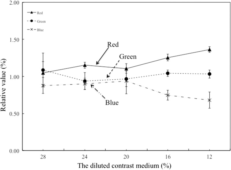
Figure 4.
FD-PBV/MR-CBV ratio in the RGB scale. MR-CBV image represents the amount of contrast medium inflowing and outflowing. Thus, a laterality differential of the CBV was not seen for either cerebral hemisphere, as shown in Figure 1. According to contrast values for comparison of the right–left CCA, we confirmed that no laterality differential was seen between CCA and VA during aortic artery flow. FD-PBV is a color-coded image that represents the quantity of the contrast medium. relative blood flow. Red = high relative blood flow. Blue = low relative blood flow. To compare FD-PBV with MR-CBV could enable a relatively fixed-quantity evaluation. Abbreviations: CBV, cerebral blood volume; FD, flat-panel detector; PBV, parenchymal blood volume; RGB, red/green/blue

