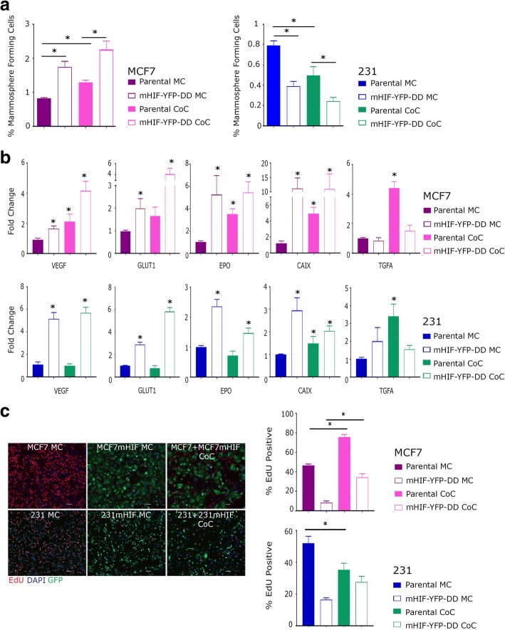
Fig. 3.
Following 48 h in the presence of TMP in either mono-culture (MC) or co-culture (CoC), mHIF-YFP-DD and parental cells were plated into mammosphere culture or used to make RNA for qRT-PCR. a Mammosphere formation increased or decreased in the mHIF-YFP-DD expressing lines as expected in MCF7 and 231 cells respectively. Interestingly, mammosphere forming cell number was also affected in the both parental cell lines following co-culture. b Gene expression changes were seen within the HIF1-alpha inducible cells as expected. Changes were also visible, however, in the parental cells grown in co-culture. c Representative photomicrographs showing EdU stain in mono- and co-culture, Green anti-GFP, Blue DAPI and Red EdU. Scale bar equal to 25 μm. Proliferation, assessed by counting cells positive for EdU, was increased in mHIF-YFP-DD and parental cells cultured in co-culture

