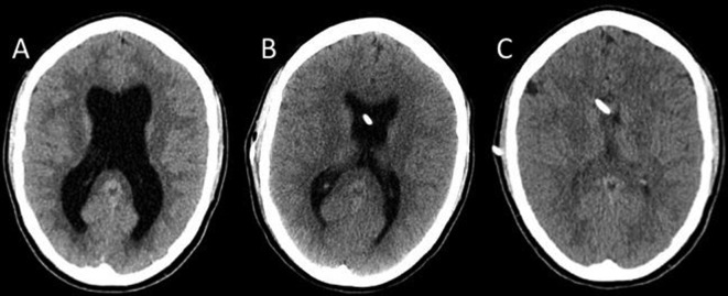
Figure 1.
Non-contrast enhanced CT scan. Images in transverse plane at the level of the lateral ventricles obtained before ventriculoperitoneal shunt (VPS). (A) Ventriculomegaly. (B and C) Increasing regression of ventriculomegaly 1 week (B) and 1 year (C) after placement of VPS, respectively. VPS catheter is seen in (B) and (C).

