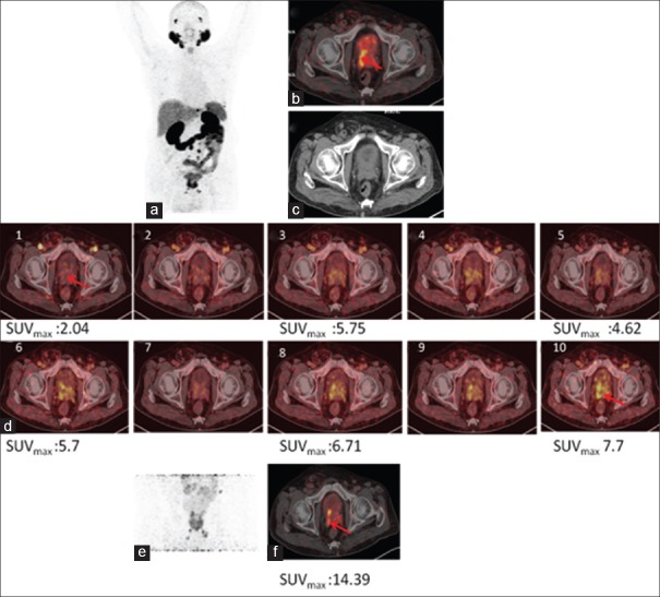
Figure 4.
68Ga-PSMA-11 positron emission tomography/computed tomography in a prostate cancer patient with biochemical recurrence. Maximum intensity projection (a), fused positron emission tomography/computed tomography (b), and computed tomography (c) of whole-body positron emission tomography 60 min p.i. showing uptake in pathologic lesion. (d) Dynamic images showing progressively increasing uptake in prostatic lesions whereas urinary bladder activity remains insignificant. (e) Showing prostatic lesion maximum intensity projection and axial section (f) on postdiuresis image

