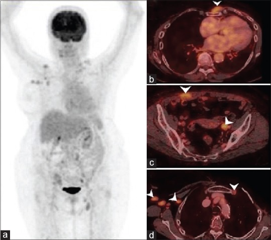
Figure 1.

(a) 18-fluorine fluoro-deoxy-glucose positron emission tomography/computed tomography maximum intensity projection image showing tracer uptake in the right axillary, left supraclavicular, left common iliac and left inguinal regions. Focal tracer uptake in right shoulder corresponds to degenerative changes in the acromioclavicular joint. (b-d) Axial fused positron emission tomography/computed tomography images showing mild 18-fluorine fluoro-deoxy-glucose uptake in subcutaneous nodule in the anterior chest wall, deposit in right rectus abdominis muscle, left external iliac, right axillary, subpectoral, and left internal mammary nodes (arrow heads)
