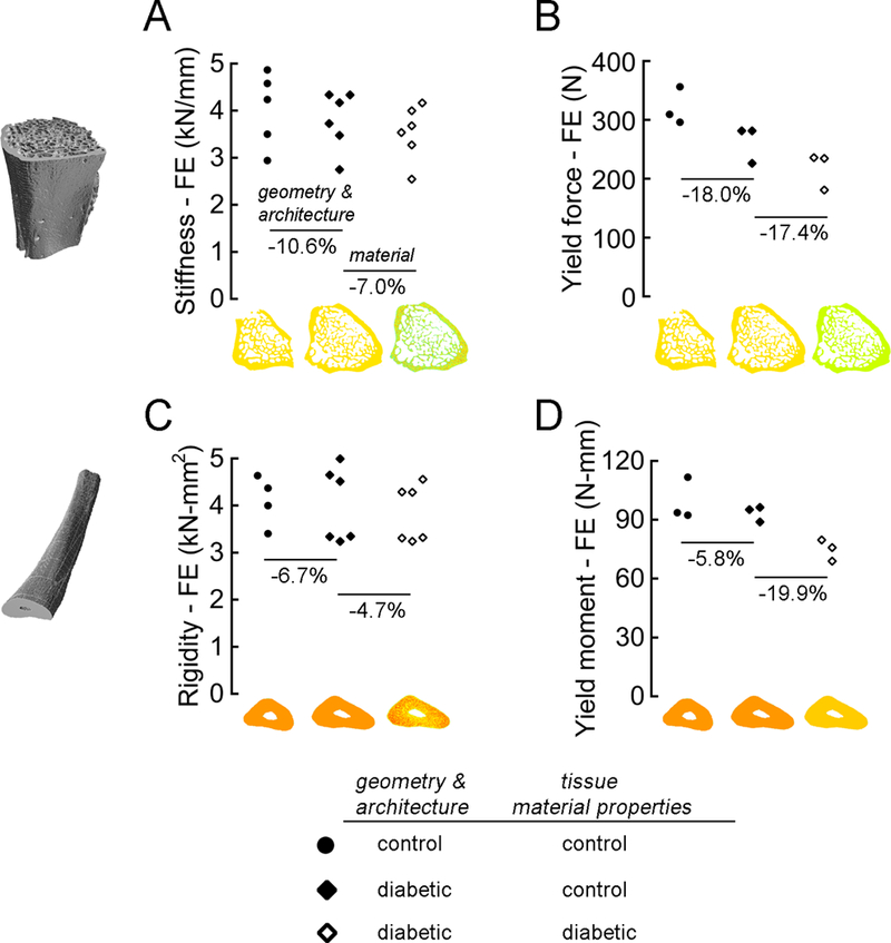
Figure 4.

High-resolution finite element analysis of the vertebrae (A & B) and ulnae (C & D) was used to estimate the relative roles of diabetes-induced deficits in bone geometry and architecture vs. material properties. Vertebral behavior was evaluated in compression; unlar mid-shaft behavior was evaluated in 3-point bending. For the elastic biomechanical properties (A & C), models of the bones from lean control rats and those with diabetes were assigned either the average tissue modulus of the bones in the control group or the specimen-specific tissue modulus derived from the micro-CT-based measurements of tissue mineral density. For the biomechanical properties at yield (B & D), models of the bones were assigned either the average tissue modulus and tissue yield strain of the bones in the control group or the specimen-specific tissue modulus and yield strain measured from the SAXS experiments.
