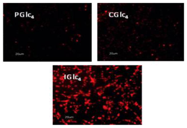
Figure 27.

Fluorescence microscopy of K:Molv NIH 3T3 cells treated with 2.5 μM PGlc4, CGlc4, and IGlc4. K:Molv NIH 3T3 cells were incubated for 20 h with porphyrinoids, followed by removal of unbound dye from the cell culture by repeated rinsing with PBS, and the cells were imaged under identical microscope settings and not enhanced; magnification 10×. Reproduced with permission from ref 234. Copyright 2010 American Chemical Society.
