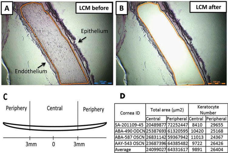
Figure 1.

Isolation of human corneal stroma by laser capture microdissection (LCM). The 9–10 mm corneal tissues from the equator were collected and cryopreserved. Also, 20 μm sections were made for LCM. (a, b) The representative images of corneal sections that were cut before and after LCM. (c) Diagram defining central and peripheral corneal stroma. (d) The data summary of keratoctyes that were captured by laser from central and peripheral corneal stroma. Scale bar: 500 μm.
