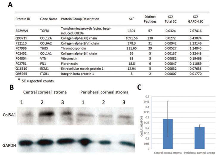
Figure 3.

The cornea stroma genes whose protein expressions were confirmed. Corneal stroma without endothelium and epithelium were subjected to LC/MS/MS proteome analysis and western blotting. (a) Proteomics analysis revealed nine proteins matching our gene findings. Among them, four were known genes and the other five were newly identified in our study. (b) Western blotting showed the positive staining for COL5A1 with a band of about 184 kD size both in central stroma and peripheral stroma samples. GAPDH was used as loading control. (c) Densitometric analysis of the mean relative intensity (n = 3) for Col5A1 normalized by GAPDH showed no significant difference between the central and peripheral corneal stroma (p = 0.55).
