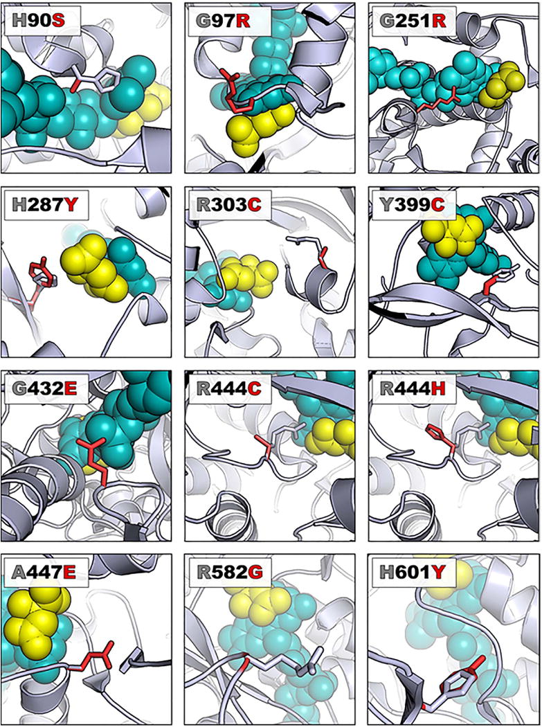
Figure 3. Variants involving the active site cause loss of Sdh1 function.

A ribbon representation of Sdh1 (E. coli model PBD file 2WP9) is shown with WT protein carbons colored gray and the variant carbons colored red. The FAD is shown as teal-colored spheres while the dicarboxylic acid substrate (succinate analog) is depicted with yellow-colored spheres. In each case, the view has been rotated so that residues of interest are clearly observed. References detailing structural implications of each variant can be found in Table 2.
