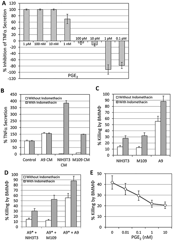
Fig. 2.

(A) Effect of PGE2 on TNFα secretion by LPS-stimulated macrophages. Hundred thousand macrophages that have been seeded in each well of a 96-flat bottomed tissue culture plate, were exposed to 1 μg/ml LPS in the absence or presence of various concentrations of PGE2 as indicated. The amount of TNFα secreted was analyzed 24h later. 1 nM corresponds to 352.4ng/ml PGE2. p<0.05 for 1 nM-1 μM and 0.1–1 pM PGE2 versus control medium. (B) Indomethacin treatment of N1H3T3 and M109 cells abolished the macrophage inhibitory effect of conditioned medium. Hundred thousand macrophages were incubated in control medium or conditioned media from untreated or indomethacin (50 μM, 24h)-treated M109, N1H3T3 and A9 cells in the presence of 1 μg/ml LPS, and the amount of TNFα secreted was determined 24h later. The data are presented as relative TNFα secretion by macrophages under each treatment condition in comparison to that of control medium. p<0.05 for N1H3T3 CM and M109 CM in the absence of indomethacin versus control and A9 CM; and p<0.05 for N1H3T3 CM and M109 CM in the presence versus in the absence of indomethacin. (C) Indomethacin sensitized the resistant tumor cells to macrophage cytotoxicity. N1H3T3, M109 and A9 cells were incubated with macrophages in the presence of 1 μg/ml LPS with or without 50 μM indomethacin for 3 days, and the extent of tumor cell killing determined. p < 0.05 for cells in the presence versus in the absence of indomethacin. (D) The presence of resistant cells prevented macrophage cytotoxicity on sensitive cells that could be reversed by indomethacin. Five thousand [3 H]-thymidine-labeled A9* cells were addedto 1 × 10⁵ macrophagesthat have been pre-incubated with 5 × 10³ unlabeled N1H3T3, M109 or A9 cells for 24 h. The extent of A9 cell killingwas determined after 3 days co-incubation. p<0.05 forkilling of A9 cells inthe presenceversus inthe absence of indomethacin. (E) PGE2 suppressed macrophage-mediated cytotoxicity of A9 cells. PGE2 was added at 10pM, 100pM, 1 nM and 10 nM to the co-culture of A9 cells and macrophages in the presence of 1 μg/ml LPS, and the extent of cell killing determined 3 days later. p<0.05 forcell killing in the presence versus in the absence of PGE2.
