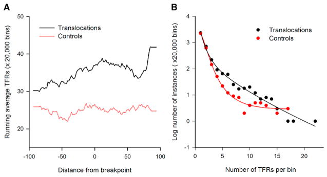
Figure 1. H-DNA TFRs Are Enriched at Translocation Breakpoints in Human Cancer Genomes.
(A) Running average of triplex-forming repeats (TFRs) in the cancer translocation breakpoint (black) and control (red) datasets, whose center loop positions are located at each base along the ±100 bp flanking the cancer translocation breakpoints or control sites.
(B) Distribution of number of bins/20,000 bins each containing various numbers of TFRs in cancer translocation breakpoint (black) and control (red) datasets.

